Postoperative ct scan is most accurate way to determine posterior wall accuracy of reduction which has greatest correlation with clinical outcome. This injury is caused by a blow to either the side or front of the knee and often occurs as a dashboard injury accompanied by a fracture of the femur.
Acetabulum Fracture Imaging: Practice Essentials ... from img.medscapestatic.com
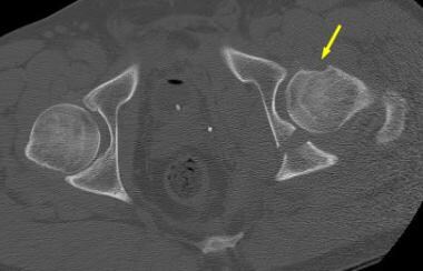
Approximately 30% of pelvic fractures seen on ct can be missed on plain films (resnik et al. Ebraheim's educational animated video describes fracture types of the acetabulum. The acetabulum is formed by an anterior and a posterior column of the bone.
Approximately 30% of pelvic fractures seen on ct can be missed on plain films (resnik et al.
Compounded by the location in a region challenging. For example, the bone can break straight across the socket or shatter computed tomography (ct) scans. Overview of pelvic fractures, acetabular fractures, treatment options and complications. 7, 8, 9, 10, 11 the anatomy of the acetabulum and computed tomography (ct) images of fractures. And medial displacement of the femoral head. The hip joint is a ball and socket joint where the femoral head is. Displaced fx with roof arcs > 45 degrees in ap and judet views or > 10 mm on axial ct cuts. 1.acetabular fractures,imaging and managementpresented by : Accurate characterization of acetabular fractures can be difficult because of the complex fracture patterns on radiography are correlated with ct, including multiplanar reconstruction and 3d. Ebraheim's educational animated video describes fracture types of the acetabulum. Postoperative ct scan is most accurate way to determine posterior wall accuracy of reduction which has greatest correlation with clinical outcome. Acetabular fractures are a type of pelvic fracture, which may also involve the ilium, ischium or acetabular fractures are uncommon. The reported incidence is approximately 3 per 100,000 per year. This view shows the characteristic comminution of the medial acetabular wall. Because of the complex anatomy of the pelvis, a ct scan is. Once the acetabular fracture is classified, appropriate therapy may be planned and implemented. Compounded by the location in a region challenging.