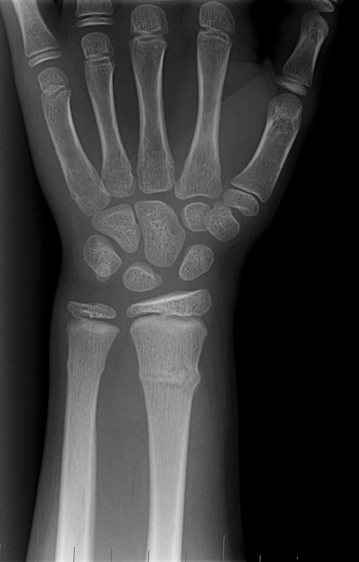
Radiology department of the rijnland hospital in leiderdorp, the netherlands. Forearm radiograph shows a torus (buckle) fracture of distal radius and ulna is most common fracture in lower forearm of young.
Fracture Wrist - Fracture Treatment from img.medscape.com
Buckle (torus) fractures in childhood are very common, and most assume a typical configuration 1 department of radiology, the university of texas medical branch, galveston, texas, usa. Buckle fracture greenstick fracture periosteum and cortex are fractured on one side and buckled on other side Torus fracture (= buckle fracture):
Buckle or torus fractures are simple axial load induced compression fractures which manifest as buckling, kinking, or notching of the cortex.
· fractures of the distal radius and/or ulna should be seen by the pcp or orthopedic clinic within 5 distal radius fractures in children: There is a buckle fracture at the ulnar aspect of the base of the fourth proximal phalanx. Care guide for buckle fracture. A buckle fracture is a break that does not go completely through the bone. Neurovascular complications are rare but should be assessed. When obtaining a radiograph of an injured limb, it is also considered good practice to obtain a radiograph of. Posted on march 6, 2020 by edg17001. One side of the bone buckles (bulges) when pressure is applied to the other side of the bone. Case contributed by assoc prof frank gaillard ◉ ◈. Suzanne o'hagan 18 may 2012. Whаt doеs а torus frаcturе look likе? They happen when one side of the bone buckles, or bends, but doesn't break all the way through. Incomplete fracture in children are: Incomplete compression fracture at metaphysis of distal radius in which the cortex is disrupted on compression (concave) side of injured bone, and remains intact on tension (convex). It is often caused from falling on the hand. · fractures of the distal radius and/or ulna should be seen by the pcp or orthopedic clinic within 5 distal radius fractures in children: Torus fracture of the first metatarsal radiology case.