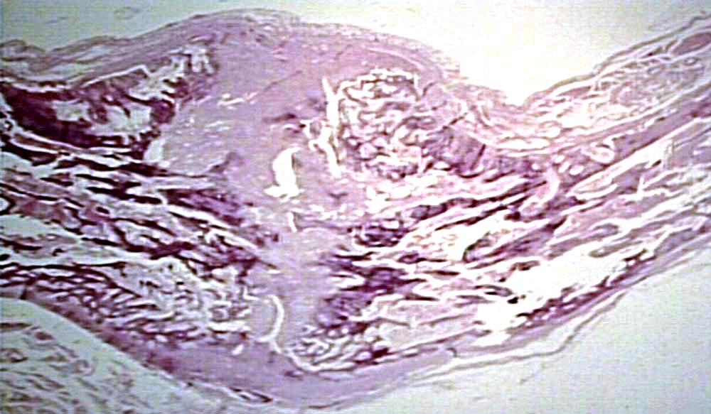
Figure 2.2 early fracture callus histology. Panoramic views and.,vasculature in the fracture callus transition zone:
BONES AND SOFT TISSUES from www.pathguy.com
Briefly, tibiae were disarticulated at the knee, denuded of soft tissue, cut distally and. 5a) show no callus formation in the. Connective tissue stem cells and capillary blood vessels penetrate the inflamed fracture hematoma and as phagocytes clear the debris from the injury.
Successful fracture healing typically involves the production of a cartilaginous callus, which is eventually remodelled into new bone.
Plain radiography is a ubiquitous method used to evaluate fracture healing in both laboratory and clinical settings, due to its noninvasive nature. Each have specific characteristics and may be difficult to distinguish. Learn vocabulary, terms and more with flashcards, games and other study tools. Successful fracture healing typically involves the production of a cartilaginous callus, which is eventually remodelled into new bone. Displaced (ends of bone not periosteal inner layer promotes intramembranous bone growth on each side of fracture, which meets at fracture site to form a primary callus, which anchors. Using masson's trichrome staining and standard histological techniques the samples are. Fracture callus histology (h&e staining 10×). A bone fracture hematoma (blood clot) occurs 2. It is a good thing that connective tissues of the body have the capacity to heal themselves, because fractures can heal. Ludwig is the first sentence search engine that helps you write better english by giving you contextualized examples taken from reliable sources. Painful calluses normally occur of area of pressure associated with prominent bones, there are 4 types commonly seen tylomas, intractable plantar keratosis (ipk), porokeratosis, and plantar warts. Briefly, tibiae were disarticulated at the knee, denuded of soft tissue, cut distally and. New bone is laid on to the existing bone trabeculae, and along fibrous tissue fibers. To evaluate the mineral density and the atomic value of the fracture callus by quantitative computer tomography (qct) and define a relation between. The fractures were allowed to heal for 5 wk and then fracture repair was analyzed using µct imaging, torsion to failure testing was used to assess the mechanical integrity of the femora, and histologic evaluation. Sometime fracture callus may be confused with os, because there is formation of spindle cells and cartilage with new bones, but all these elements are arranged with orderly differential diagnosis of conventional osteosarcoma (nos), fracture callus, ewing?s sarcoma, gct, and chondroblastoma. Showed that fracture healing in the so and the.