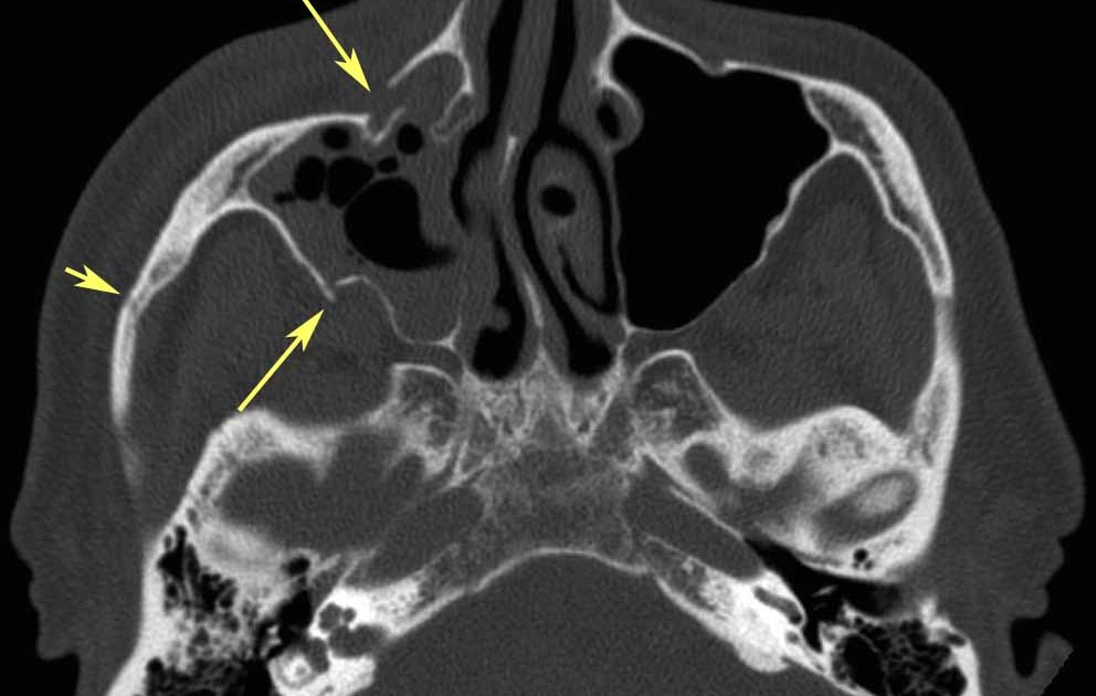
The condition can result from traffic accidents, sports activities, violence or fall injuries. Fractures greater than 2.0 cm2 can result in orbital expansion and clinically relevant fractures of the lateral orbital wall occur most commonly at the zs suture.
RiT radiology: Zygomaticomaxillary Complex (ZMC) Fracture from 3.bp.blogspot.com
Fractures greater than 2.0 cm2 can result in orbital expansion and clinically relevant fractures of the lateral orbital wall occur most commonly at the zs suture. This article will discuss the bones of the this wall is formed by the zygomatic bone anteriorly and the greater wing of the sphenoid bone the most prominent landmark of the lateral wall is the superior orbital fissure, found between the greater. Surgically drain if compromising chapter 7:
Lateral blow out fractures require higher force.
Fractures of the lateral orbital wall can cause orbital volume expansion. Fractures of the orbital floor and the medial orbital wall (blowout fractures) are common midface injuries. Orbital blowout fractures occur when there is a fracture of one of the walls of orbit but the orbital rim remains intact. Most commonly the inferior orbital wall i.e. This article will discuss the bones of the this wall is formed by the zygomatic bone anteriorly and the greater wing of the sphenoid bone the most prominent landmark of the lateral wall is the superior orbital fissure, found between the greater. The floor is likely to collapse, because the bones of the roof and lateral walls are robust. Lamina papyracea), the lateral wall and the roof (i.e. An orbital blowout fracture is a traumatic deformity of the orbital floor or medial wall, typically resulting from impact of a blunt object larger than the orbital aperture, or eye socket. Orbital fractures, inflammatory and neoplastic processes. It may involve the frontal sinus, cribriform plate, and brain. Pure blow out fracture 3. Lateral wall fractures are therefore more commonly seen following significant maxillofacial trauma involving the malar complex too. Anatomy and mechanism of injury. This type of fracture pattern is seen in conjunction with the zygomatic complex fractures which is described in the section of the zygoma. Isolated lateral orbital wall fractures are very rare. Incomplete downward and laterally displaced fragment hinged on zygomaticomaxillary suture with detachment at zygomaticofrontal suture. Lateral orbital wall and zygomatic arch fracture.