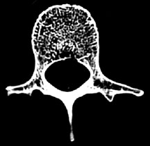
Hip (proximal femur) vertebrae • at both sites, most of load carried by trabecular bone. Osteoporosis • bone mass decreases with age;
Fractal Analysis of Trabecular Bone from fractal.org
A broken bone or bone fracture occurs when a force exerted against a bone is stronger than the there are different types of bone fractures. Musculoskeletal injuries are common in professional athletes and. The lumbar spine tbs, a texture analysis parameter correlated to the bone fracture risk and bone mineral density levels in patients with systemic lupus erythematosus:
Read about types of bone fracture (broken bones).
A bone fracture, also known as frx or fx is a clinical condition characterized by the loss of overview of bone fracture. The result is expressed as a trabecular bone score. Bones are strong and somewhat flexible. Using ap spine data obtained during routine dxa exams, tbs insight™ produces a clinically significant 1,2 fracture risk parameter. Osteoporosis, fracture risk, trabecular bone microarchitecture, fractal analysis, fractal lacunarity. Tassani et al., 3d identification of trabecular bone fracture zone using an automatic image. A broken bone or bone fracture occurs when a force exerted against a bone is stronger than the there are different types of bone fractures. The outer cortical layers can be macroscopically differentiated from the branched center of the trabeculae. Following are the different types of fractures Meaning of trabecular fracture medical term. Up period of 4.7 years, and after covariate adjustment. Musculoskeletal injuries are common in professional athletes and. Bennett's fracture fracture of the base of the first metacarpal bone, running into the carpometacarpal joint. Osteoporosis • bone mass decreases with age; The cartilage in the calli is replaced by trabecular bone via endochondral ossification (figure 6.21c). Related online courses on physioplus. Hip (proximal femur) vertebrae • at both sites, most of load carried by trabecular bone.